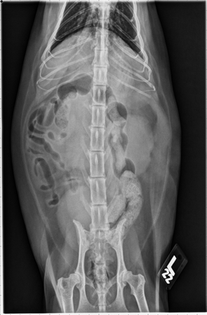
Abdominal radiographs show both kidneys are enlarged with rounded and irregular margins, right more severe than the left. The spleen is mildly enlarged with rounded margins. An oval soft tissue opacity is present ventral to S1 with ventral displacement of the cranial rectum. Portion of the thoracic cavity that is visible appears unremarkable.
Dorsoventral view:
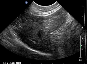
Abdominal ultrasound shows both kidneys are enlarged with irregular margins and perirenal hypoechoic rims. Both renal cortices are also mottled in appearance with ill-defined multiple hypoechoic nodules. A small amount of aggregation of gravity-dependent hyperechoic sediment is present within the urinary bladder (not pictured).
Image of right kidney: