Cerebrospinal nematodiasis due to Parelaphostrongylsus tenuis occurs in South American camelids (eg, llamas and alpacas) in areas of the United States where populations overlap with white-tailed deer (Odocoileus virginianus), who are the definitive host. Rarely causing clinical signs in the definitive host, P tenuis infection creates severe neurologic problems due to aberrant migration in dead-end hosts, such as South American camelids and other cloven hoof stock. Infestation occurs after ingestion of an intermediate host gastropod (slug or snail) containing third-stage larvae. The larvae migrate from the gastrointestinal tract to the spinal cord and continue cranially, creating a destructive tract in the spinal cord on the way to the brain. Clinical signs are commonly asymmetric pelvic limb neurologic deficits that progress cranially. The disease often goes undetected until the animal becomes recumbent. Definitive diagnosis is based on postmortem necropsy findings (shown below) when the nematodes are found in histologic sections. Antemortem diagnosis usually relies on a combination of neurologic exam findings, confirming exposure to white-tailed deer, increased eosinophils in cerebrospinal fluid (eosinophilic pleocytosis), and response to treatment with anthelmintic and anti-inflammatory drugs. Commercial kits are not available for testing. Fecal evaluation is not valuable because the first-stage larvae do not develop in aberrant hosts.
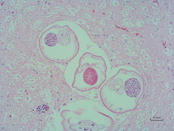
Necropsy findings: Definitive diagnosis of P tenuis infection requires finding the organisms in CNS tissue and results of molecular testing for species identification. Molecular testing is not widely available. In this case, multiple nematode cross-sections were found in the brainstem (medulla), and the spinal cord showed areas of damage attributed to migration.
Links to sections in MVM:
Nematodes Causing CNS Disease
Herd Health of Llamas and Alpacas ‐ Parasite Control
Management of Llamas and Alpacas
References
Pinn TL, Bender HS, Stokol T, Erb HN, Schlafer DH, Perkins GA. Cerebrospinal fluid eosinophilia is a sensitive an specific test for the diagnosis of Parelaphostrongylus tenuis in camelids in the northeastern United States. J Vet Diagn Invest. 2013 Jan;25(1):54-60. doi.org/10.1177/1040638712471058
Dobey CL, Grunenwald C, Newman SJ, Muller L, Gerhold RW. Retrospective study of central nervous system lesions and association with Parelaphostrongylus species by histology and specific nested polymerase chain reaction in domestic camelids and wild ungulates. J Vet Diagn Invest. 2014 Nov;26(6):748-54. doi: 10.1177/1040638714553427